An innovative artificial intelligence model has demonstrated the capacity to identify critical cancer clues directly from tissue slides, specifically by detecting genetic weaknesses within ovarian cancer samples. This development represents a significant step towards leveraging AI for enhanced diagnostic precision and guiding personalized treatment strategies in oncology.
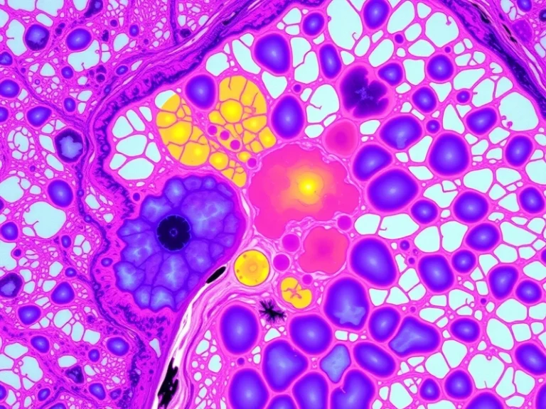
The core of this AI innovation lies in its ability to analyze complex histopathological images—microscopic views of tissue biopsies—and discern subtle patterns indicative of underlying genetic alterations. Traditional pathology relies heavily on the human eye and specialized staining techniques, often followed by separate molecular tests to identify specific genetic markers. By contrast, this AI approach aims to extract this molecular-level information directly from standard H&E (hematoxylin and eosin) stained slides, which are routinely prepared in pathology labs worldwide.
The Mechanism: Deep Learning in Histopathology
At its foundation, the AI model likely employs deep learning architectures, most commonly convolutional neural networks (CNNs), which are particularly adept at image recognition tasks. These networks are trained on vast datasets of digitized tissue slides, where each slide is meticulously annotated with known genetic statuses—for instance, whether a sample carries a specific mutation or exhibits a particular genetic deficiency. Through this extensive training, the AI learns to identify visual proxies for these molecular characteristics. These proxies might be subtle changes in cell morphology, nuclear size and shape, tissue architecture, or the distribution of immune cells within the tumor microenvironment, which are often imperceptible or difficult to consistently quantify by human observation alone.
- Image Preprocessing: Digitization of glass slides into high-resolution images.
- Feature Extraction: CNNs automatically learn relevant visual features from these images.
- Pattern Recognition: The model identifies correlations between visual features and known genetic states.
- Prediction: Based on learned patterns, the AI predicts the likelihood of specific genetic weaknesses in new, unseen tissue samples.
Why Ovarian Cancer? A Critical Need for Precision
The application of this technology to ovarian cancer is particularly impactful. Ovarian cancer is often diagnosed at advanced stages due to its non-specific symptoms, leading to poorer prognoses. Furthermore, a significant subset of ovarian cancers, particularly high-grade serous ovarian carcinoma (HGSOC), are characterized by specific genetic vulnerabilities that make them highly responsive to targeted therapies. Identifying these vulnerabilities early and efficiently is crucial for optimizing patient outcomes.
One of the most well-known genetic weaknesses in ovarian cancer involves mutations in the BRCA1 and BRCA2 genes, or more broadly, the presence of homologous recombination deficiency (HRD). HRD signifies a cell’s impaired ability to repair double-strand DNA breaks, making these cells particularly susceptible to DNA-damaging agents and therapies that exploit this repair defect. Poly (ADP-ribose) polymerase (PARP) inhibitors, such as olaparib, niraparib, and rucaparib, are a class of targeted drugs highly effective in treating ovarian cancers with BRCA mutations or HRD. However, identifying patients who will benefit from these therapies typically requires expensive and time-consuming molecular genetic testing.
AI’s Potential Impact on Diagnosis and Treatment Selection
By potentially inferring BRCA status or HRD directly from routine histopathology slides, the AI model could offer several advantages:
- Speed and Efficiency: It could provide a rapid initial screening tool, potentially flagging patients who are strong candidates for PARP inhibitors much faster than traditional sequencing methods.
- Cost Reduction: By reducing the need for universal, complex molecular testing, it could lower diagnostic costs, making precision medicine more accessible.
- Accessibility: In regions with limited access to advanced molecular diagnostics, an AI-powered histopathology analysis could democratize access to critical information for treatment planning.
- Enhanced Pathologist Workflow: The AI could act as an assistive tool, highlighting suspicious regions or providing a second opinion, thereby improving diagnostic consistency and reducing workload for human pathologists.
This innovative use of AI does not aim to replace the human pathologist but rather to augment their capabilities, providing an additional layer of data-driven insight. Pathologists would still review the AI’s findings, integrating them with their expertise and other clinical information to arrive at a comprehensive diagnosis and treatment recommendation.
Challenges and Future Directions
While promising, the path to clinical implementation for such AI models involves rigorous validation. The model must demonstrate high accuracy, sensitivity, and specificity across diverse patient populations and different pathology labs to ensure its reliability. Furthermore, regulatory approval processes will require extensive testing and clear evidence of clinical utility and safety. Issues of interpretability—understanding why the AI makes a particular prediction—are also crucial for building trust among clinicians and patients.
Future research will likely focus on refining these models, expanding their ability to detect a broader range of genetic markers, and integrating them seamlessly into existing digital pathology workflows. The ultimate goal is to move towards a future where AI-driven insights from routine tissue samples can empower clinicians to make more informed, personalized treatment decisions, ultimately improving patient outcomes in challenging cancers like ovarian carcinoma.